See Beneath the Surface with AI-Enabled Ultrasound and Inject with Confidence
Portable ultrasound, AI-powered facial assessment, training, and clinical onboarding designed to help aesthetic professionals improve precision, anatomy awareness, procedural confidence, and patient safety.
Trusted by Clinicians, Educators & Healthcare Professionals Across the UK
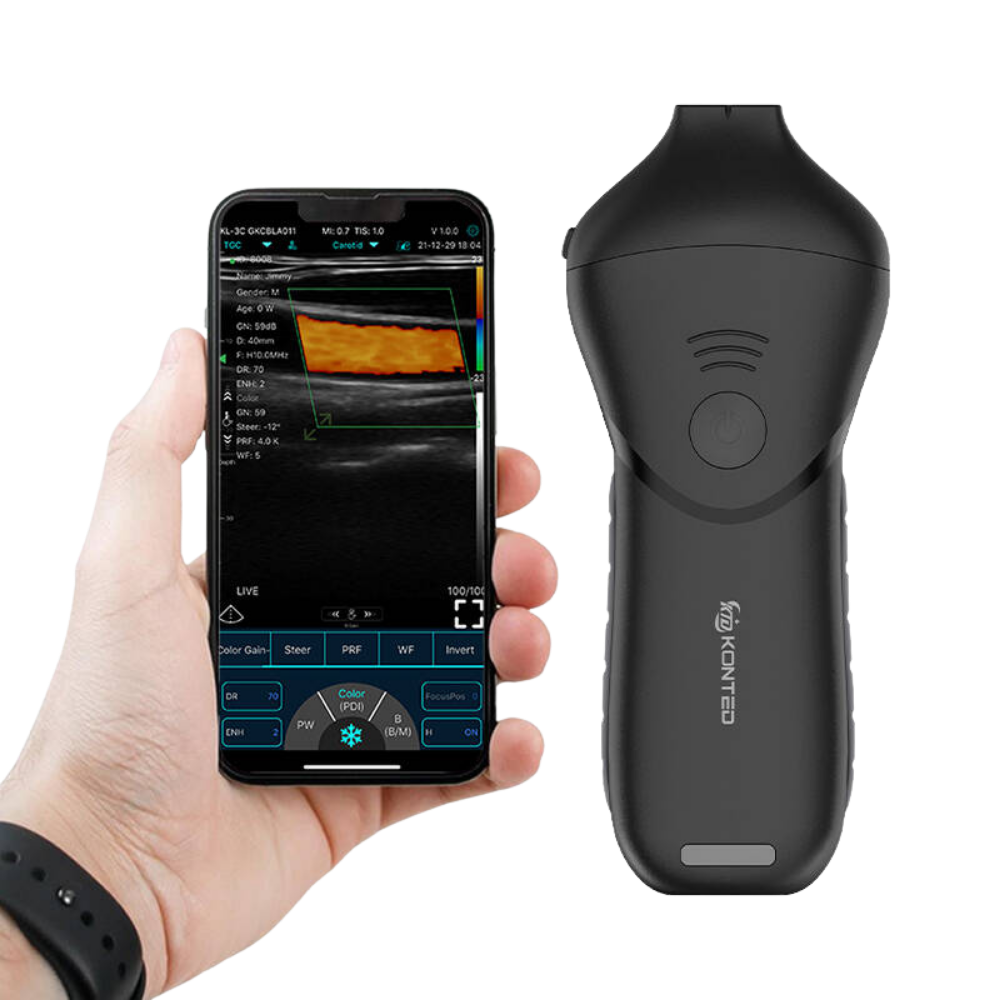
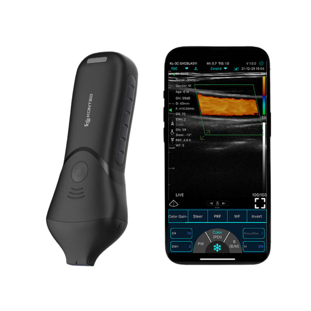
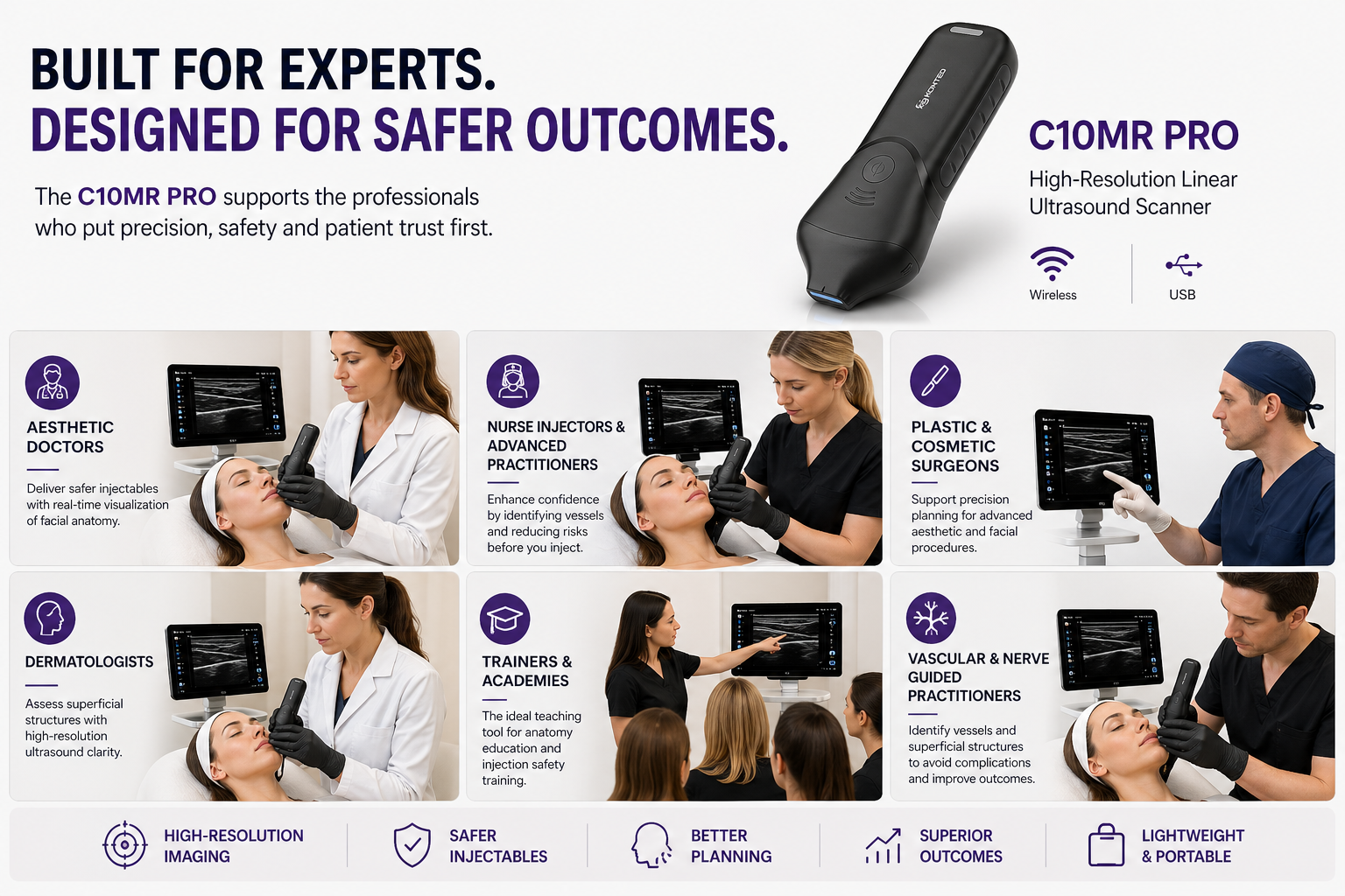
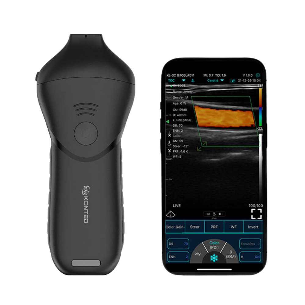
Delivering ultra-high-resolution imaging for facial anatomy, skin, vessels, filler assessment, and soft tissue, the High Linear Ultrasound Probe dual-frequency (18/24 MHz) probe is ideal for aesthetic, dermatology, vascular, and superficial MSK applications.
With USB and WiFi connectivity, it pairs seamlessly with smartphones, tablets, and laptops - providing portable, high-precision imaging without bulky equipment.
The Problem
Most Aesthetic Procedures Are Still Performed Without Real-Time Anatomical Visibility
Even experienced practitioners often rely on:
But every patient is different.
Without imaging visibility, practitioners may face:
Modern aesthetic practice is evolving toward:
data-driven precision.
The Solution
The AI-Enabled Ultrasound-Guided Aesthetics Starter System
A complete adoption pathway designed to help clinics integrate ultrasound confidently into everyday aesthetic and skin practice.
What’s Included:
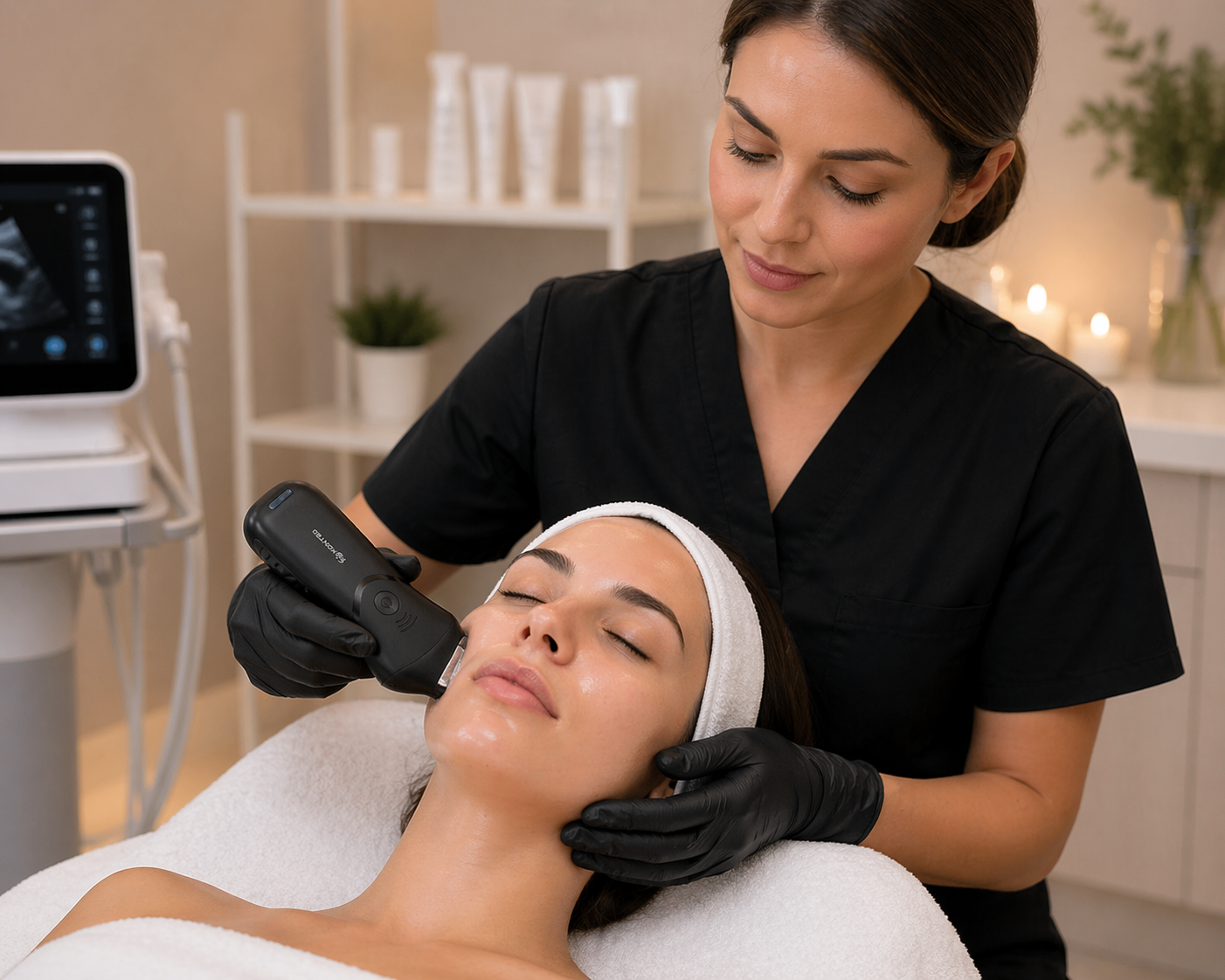
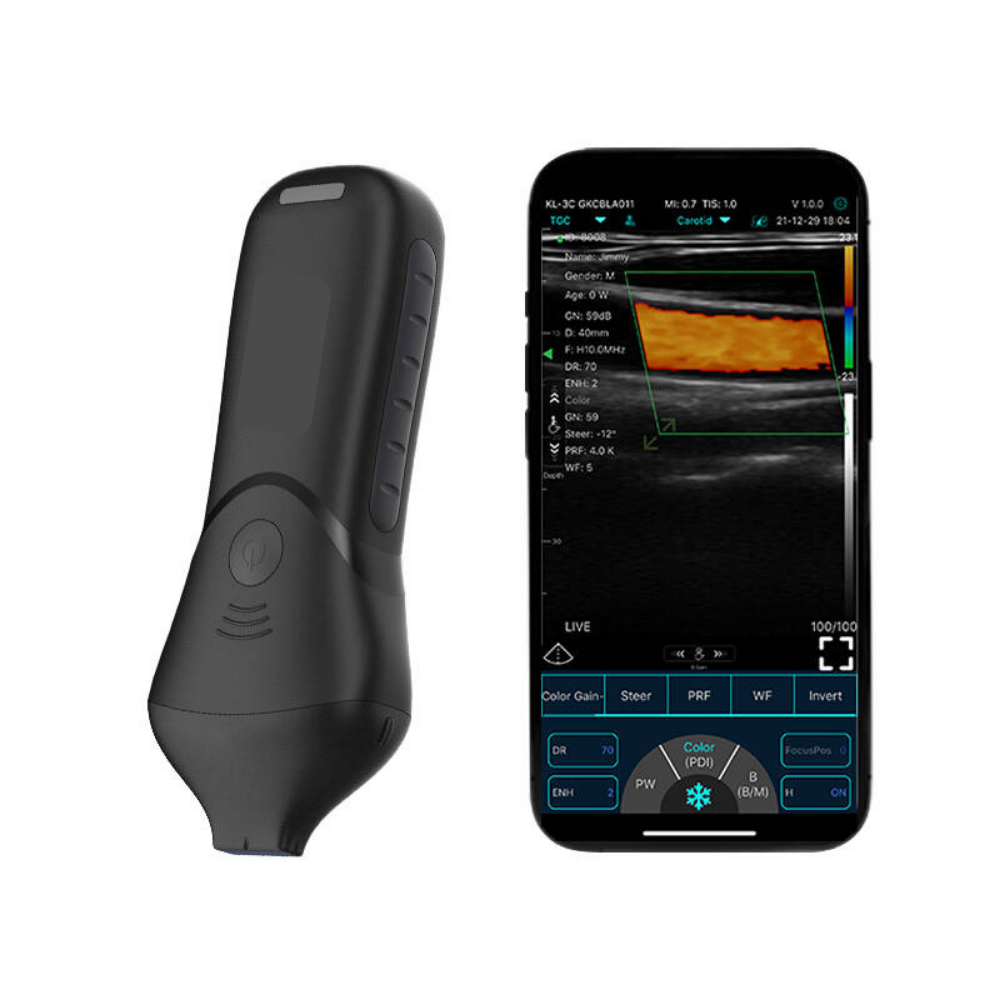
Portable AI-Enabled Ultrasound Scanner
High-resolution handheld ultrasound with advanced imaging and facial assessment capabilities.
Training & Clinical Onboarding
Structured onboarding and ultrasound-guided aesthetics education designed for real-world clinical use.
AI Facial Assessment & Skin Analysis
AI-supported full-layer facial and skin evaluation including:
Injection Guidance Support
Improve visibility during procedures with enhanced anatomical awareness and real-time imaging support.
Ongoing Clinical Support
Implementation guidance, adoption support, and optional mentorship/community access.
The issue isn’t just access to devices - it’s the ability to use them confidently when it matters most.
Flexible Finance Options Available
Spread the investment cost of your diagnostic equipment with flexible business finance options available for eligible organisations.
Flexible finance options may be available for equipment, onboarding, training, and implementation support, subject to status and approval.
Why Clinics Are Moving Toward Ultrasound-Guided Aesthetics
1. Improve Practitioner Confidence
Gain deeper anatomical insight and procedural visibility.
2. Enhance Patient Safety
Support safer treatment planning and injection precision.
3. Differentiate Your Clinic
Position your practice at the forefront of advanced aesthetic medicine.
4. Build Long-Term Patient Trust
Improve consultations through data-driven visual assessment.
5. Create a More Premium Clinical Experience
Integrate advanced imaging technology into patient journeys.
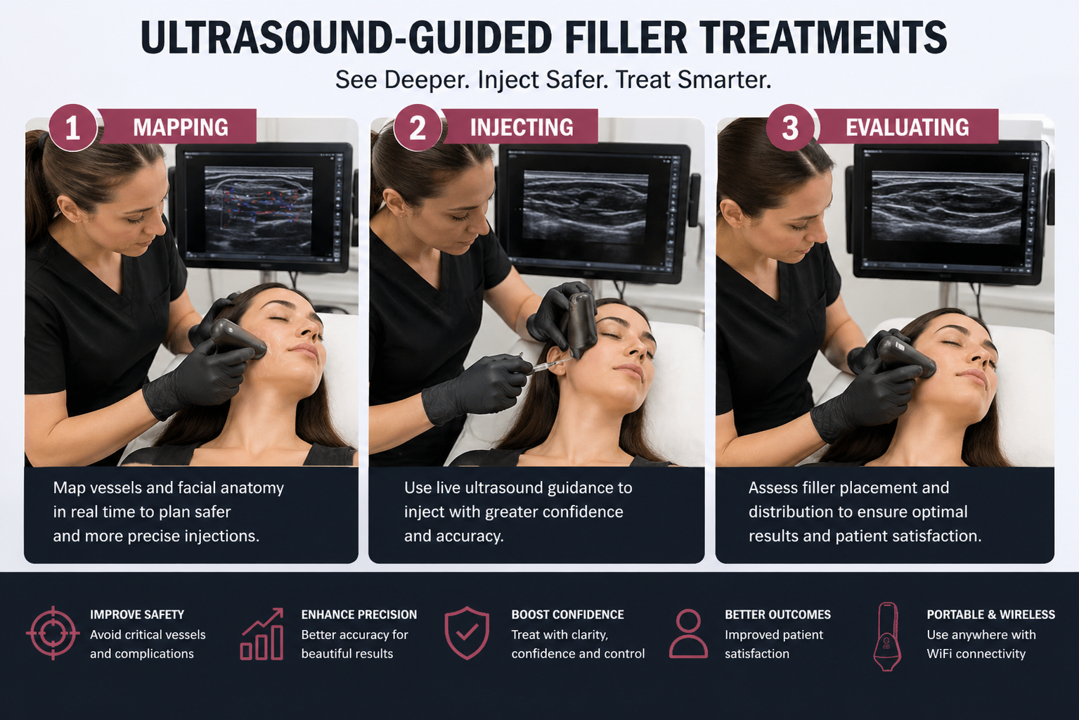
See More. Inject Safer. Treat Smarter.
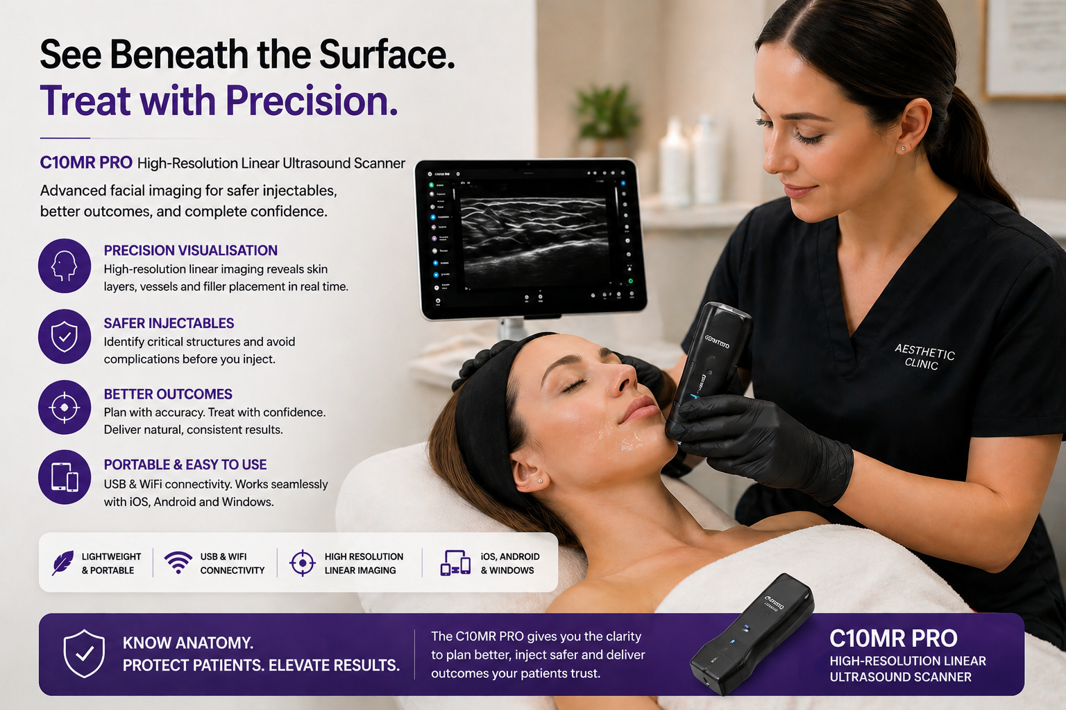
5 Key Benefits of MR PRO
1. Ultra-High Resolution Facial Imaging
With 18/24 MHz frequency, the scanner delivers exceptional detail for superficial structures such as skin layers, vessels, filler deposits, and facial anatomy.
2. Supports Safer Aesthetic Treatments
Helps practitioners visualise anatomy before or during procedures, supporting safer filler placement, complication management, and more confident decision-making.
3. Lightweight & Highly Portable
At only 155g, the probe is easy to handle during treatments, training sessions, or mobile clinic work.
4. Wireless & Multi-Device Compatible
Connects via Wi-Fi or USB and works with Android, iOS, and Windows, allowing flexible use across tablets, phones, and computers.
5. Waterproof & Clinic-Ready Design
With IP68 waterproof protection, the device is durable, hygienic, and suitable for busy professional clinical environments.
Who Is It For?
Designed for aesthetic professionals who prioritise precision, safety, and superior patient outcomes, the C10MR PRO helps practitioners see facial anatomy in real time before they treat. Ideal for clinics and clinicians who want to elevate standards, reduce risk, and deliver more confident injectable results.
Real Ultrasound Imaging Examples
See how the High Linear Ultrasound Probe helps visualise superficial facial vascular anatomy in real time. These example scans demonstrate clear imaging of key facial vessels, supporting safer treatment planning, improved anatomical understanding, and greater practitioner confidence during advanced aesthetic procedures. Ideal for clinics prioritising precision, patient safety, and evidence-led practice.

Angular artery
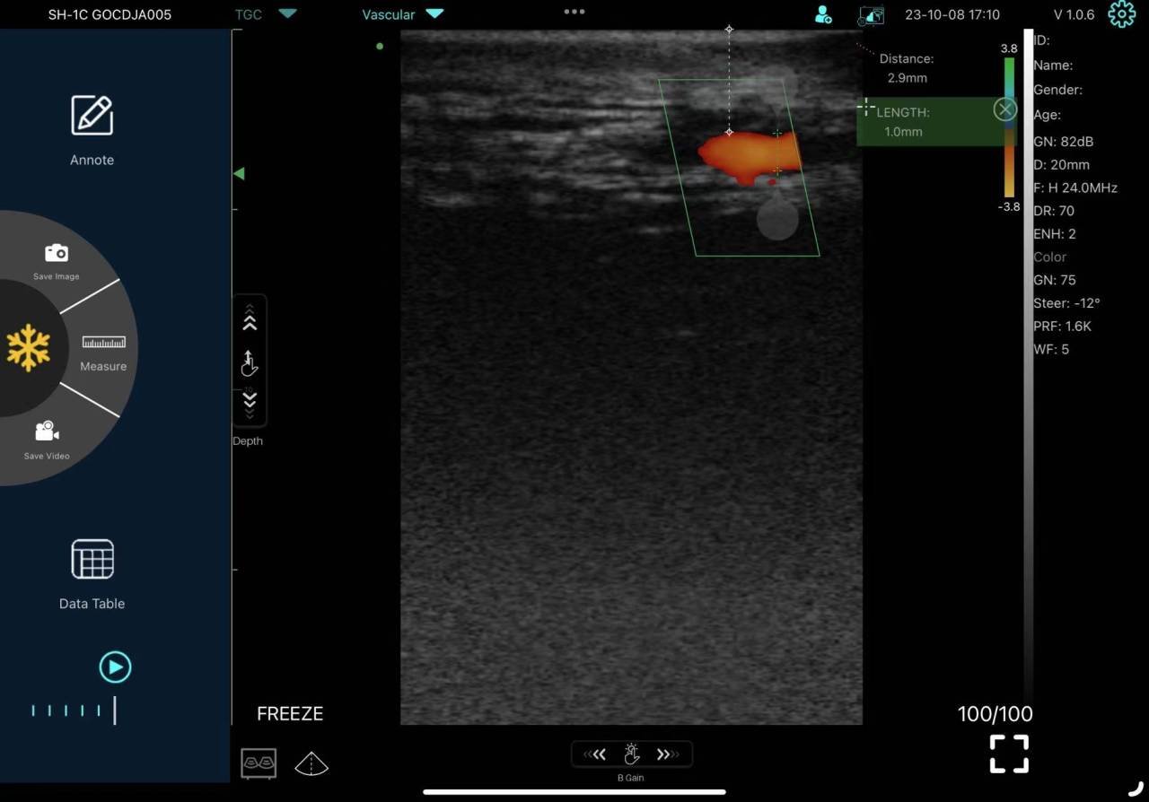
Anterior deep temporal artery
Clinical & Aesthetic Use Cases
The High Linear Ultrasound Probe supports facial aesthetic and superficial imaging applications, from treatment planning and anatomy assessment to complication management and education. Its portability and image clarity help practitioners make safer, more informed decisions in real time.
Facial Filler Mapping
The scanner can help practitioners identify existing dermal filler placement, depth, and distribution beneath the skin. This is particularly useful for clients who have had previous treatments elsewhere or cannot remember what product was used. By visualising filler locations, practitioners can plan safer retreatments, corrections, or dissolving procedures with greater confidence.
Vascular Assessment
Facial anatomy contains important blood vessels that vary between individuals. Ultrasound imaging can help locate superficial vessels in treatment zones such as the lips, nose, cheeks, temples, and tear trough area. This may support safer treatment planning by helping practitioners avoid high-risk areas and reduce the likelihood of vascular complications.
Facial Anatomy Education & Training
The scanner is a powerful educational tool for injectors, doctors, nurses, and aesthetic trainees. Users can observe muscles, vessels, tissue layers, and filler placement in real time, helping bridge the gap between textbook anatomy and live patient anatomy. This supports better technique development and deeper anatomical understanding.
Lip Filler Procedures
The lips are highly vascular and delicate treatment areas. Ultrasound can help assess tissue structure, previous filler, uneven product placement, or nodules before retreatment. This may improve planning for safer and more precise lip enhancement procedures.
Managing Filler Complications
When swelling, lumps, asymmetry, migration, or suspected vascular compromise occurs, ultrasound can provide valuable real-time information. It may help determine whether symptoms relate to filler presence, fluid, inflammation, or tissue changes. This can assist in deciding the most appropriate next steps, such as targeted dissolving or referral.
Tear Trough Treatments
The tear trough area is anatomically complex, containing vessels, thin skin, and sensitive structures. Ultrasound can support assessment of previous filler, oedema, migration, and tissue quality before additional treatment or dissolving. This can help practitioners make more informed decisions in a high-risk area.
Temple & Midface Treatments
The temples and midface can contain significant vascular structures and varying tissue depth. Ultrasound may help assess anatomy prior to filler placement, especially in advanced treatments where precision is important. This can support safer treatment planning and confidence.
Skin Lesions, Lumps & Swelling Assessment
The scanner may be used to review superficial skin irregularities, swelling, nodules, or palpable lumps. While not a replacement for formal medical diagnosis, it can help practitioners better understand superficial tissue characteristics and guide appropriate referral where needed.
Pre-Treatment Consultation Tool
Using ultrasound during consultations can add a premium diagnostic element to the patient journey. It helps explain anatomy, previous filler findings, or treatment limitations visually, improving trust, education, and informed consent discussions.
Premium Clinic Differentiation
Offering ultrasound-guided assessment can position a clinic as safety-led, modern, and clinically advanced. This may help attract more informed patients seeking practitioners who prioritise anatomy, complication prevention, and evidence-based care.
Specifications
(Click a feature to expand)
C10MR PRO
High Frequency Linear Probe
Android. IOS. Windows
-Power on/off
-Freeze/unfreeze
802.11n/20MHz/
2.4G/5G
Wi-Fi
USB
18/24Mhz
10-15-20-25mm
L18*W5mm
128
32
40-110Db
155x65x25mm
155g
IP68
B
B+B
B+M
Color
PW
PDI
Dual & Full screen Display => YES
Software Control => YES
B GN (Gain) => YES
Color GN (Gain) => YES
D (Depth) => YES
ENH (Enhancement) => YES
DR (Dynamic Range) => YES
H (harmonic) => YES
Focus => YES
8 TGC => YES
Mid-Line => YES
An note => YES
Page Left/Right Up/Down => YES
18~24 F/S
Aesthetics Vascular, Nerve,VesselFlow user1 user2
B: Length, Distance, Area/Circumference, Trace, Depth, Angle,
B+M: Heart Rate, Time, Distance, LVID
B+PW: Velocity, Heart Rate(2), S/D, Depth, Time.
Yes
Yes
Yes
Built-in High capacity Lition batteries
2200 mAh,
3 W (Live) / 1 W (freeze)
2h
48h Ultra-long standby
2h
Type-C USB
Wireless charging
Yes
Yes
JPEG, MP4, Dicom
Automatic / manual
100-200-500-1000 frame
Chinese, Ukrainian, German, English, Russian, France, Italian, Spanish, Portuguese (Brazil)

Experience Precision Imaging in Real Time

Flexible Finance Options Available
Spread the investment cost of your diagnostic equipment with flexible business finance options available for eligible organisations.
Flexible finance options may be available for equipment, onboarding, training, and implementation support, subject to status and approval.
Start Your Ultrasound Adoption Journey
Book a free Clinical Adoption Session to explore:
ABOUT US
Oras Medical is a specialist clinical adoption provider helping professionals, clinics, hospitals, GPs and care teams implement portable medical ultrasound solutions across the UK.
Oras Medical is a trading name of Oras Marketing Limited, Registered in England & Wales with registration number 12935403. VAT No. GB498618427. Registered address: Eastlands Court Business Centre, St. Peters Road, Rugby, Warwickshire, CV21 3QP. Email: admin@orasmedical.com, Phone: +44 (0) 1788 270 191.
SOLUTIONS
PRODUCTS
USEFUL INFO
PAYMENT INFO
Copyright 2026 Oras Medical - Privacy policy | Terms & Conditions | Disclaimer