
Use Cases:
Facial Aesthetic Ultrasound Scanner
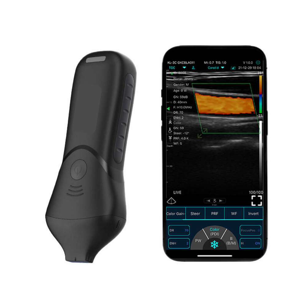
The Facial Aesthetic Ultrasound Scanner has been specifically designed to support the growing role of ultrasound within facial aesthetics. As the industry places increasing emphasis on patient safety, anatomical understanding, and evidence-based practice, ultrasound is becoming an invaluable tool for practitioners seeking greater confidence before, during, and after injectable treatments. By providing real-time visualisation of facial anatomy, clinicians can make more informed decisions while enhancing patient communication and clinical care.
Unlike traditional assessment methods that rely primarily on surface anatomy and clinical judgement, facial ultrasound enables practitioners to visualise the tissues beneath the skin. This includes facial layers, blood vessels, muscles, and existing dermal filler, helping clinicians better understand each patient's unique anatomy. Whether assessing previous treatments, planning new procedures, or investigating post-treatment concerns, ultrasound provides additional information that may support safer and more precise clinical decision-making.
The scanner can be incorporated throughout the patient journey—from the initial consultation and treatment planning to image-guided procedures, complication assessment, and follow-up reviews. It also serves as an excellent educational resource, helping practitioners develop a deeper understanding of facial anatomy while allowing patients to see and better understand their own treatment plans. This can enhance communication, strengthen informed consent discussions, and build greater trust between practitioner and patient.
As facial ultrasound continues to gain recognition across the aesthetics profession, clinics that adopt this technology are increasingly positioning themselves as safety-focused, clinically progressive, and committed to delivering high standards of patient care. The following examples demonstrate some of the many ways the Facial Aesthetic Ultrasound Scanner can support practitioners in everyday aesthetic practice. Ultrasound should always be used by appropriately trained clinicians and within their professional scope of practice.
Facial Filler Mapping
The scanner can help practitioners identify existing dermal filler placement, depth, and distribution beneath the skin. This is particularly useful for clients who have had previous treatments elsewhere or cannot remember what product was used. By visualising filler locations, practitioners can plan safer retreatments, corrections, or dissolving procedures with greater confidence.
Vascular Assessment
Facial anatomy contains important blood vessels that vary between individuals. Ultrasound imaging can help locate superficial vessels in treatment zones such as the lips, nose, cheeks, temples, and tear trough area. This may support safer treatment planning by helping practitioners avoid high-risk areas and reduce the likelihood of vascular complications.
Facial Anatomy Education & Training
The scanner is a powerful educational tool for injectors, doctors, nurses, and aesthetic trainees. Users can observe muscles, vessels, tissue layers, and filler placement in real time, helping bridge the gap between textbook anatomy and live patient anatomy. This supports better technique development and deeper anatomical understanding.
Lip Filler Procedures
The lips are highly vascular and delicate treatment areas. Ultrasound can help assess tissue structure, previous filler, uneven product placement, or nodules before retreatment. This may improve planning for safer and more precise lip enhancement procedures.
Managing Filler Complications
When swelling, lumps, asymmetry, migration, or suspected vascular compromise occurs, ultrasound can provide valuable real-time information. It may help determine whether symptoms relate to filler presence, fluid, inflammation, or tissue changes. This can assist in deciding the most appropriate next steps, such as targeted dissolving or referral.
Tear Trough Treatments
The tear trough area is anatomically complex, containing vessels, thin skin, and sensitive structures. Ultrasound can support assessment of previous filler, oedema, migration, and tissue quality before additional treatment or dissolving. This can help practitioners make more informed decisions in a high-risk area.
Temple & Midface Treatments
The temples and midface can contain significant vascular structures and varying tissue depth. Ultrasound may help assess anatomy prior to filler placement, especially in advanced treatments where precision is important. This can support safer treatment planning and confidence.
Skin Lesions, Lumps & Swelling Assessment
The scanner may be used to review superficial skin irregularities, swelling, nodules, or palpable lumps. While not a replacement for formal medical diagnosis, it can help practitioners better understand superficial tissue characteristics and guide appropriate referral where needed.
Pre-Treatment Consultation Tool
Using ultrasound during consultations can add a premium diagnostic element to the patient journey. It helps explain anatomy, previous filler findings, or treatment limitations visually, improving trust, education, and informed consent discussions.
Premium Clinic Differentiation
Offering ultrasound-guided assessment can position a clinic as safety-led, modern, and clinically advanced. This may help attract more informed patients seeking practitioners who prioritise anatomy, complication prevention, and evidence-based care.
ABOUT US
Oras Medical is a specialist clinical adoption provider helping professionals, clinics, hospitals, GPs and care teams implement portable medical ultrasound solutions across the UK.
Oras Medical is a trading name of Oras Marketing Limited, Registered in England & Wales with registration number 12935403. VAT No. GB498618427. Registered address: Eastlands Court Business Centre, St. Peters Road, Rugby, Warwickshire, CV21 3QP. Email: admin@orasmedical.com, Phone: +44 (0) 1788 270 191.
SOLUTIONS
PRODUCTS
USEFUL INFO
PAYMENT INFO

Copyright 2026 Oras Medical - Privacy policy | Terms & Conditions | Disclaimer