
Use Cases:
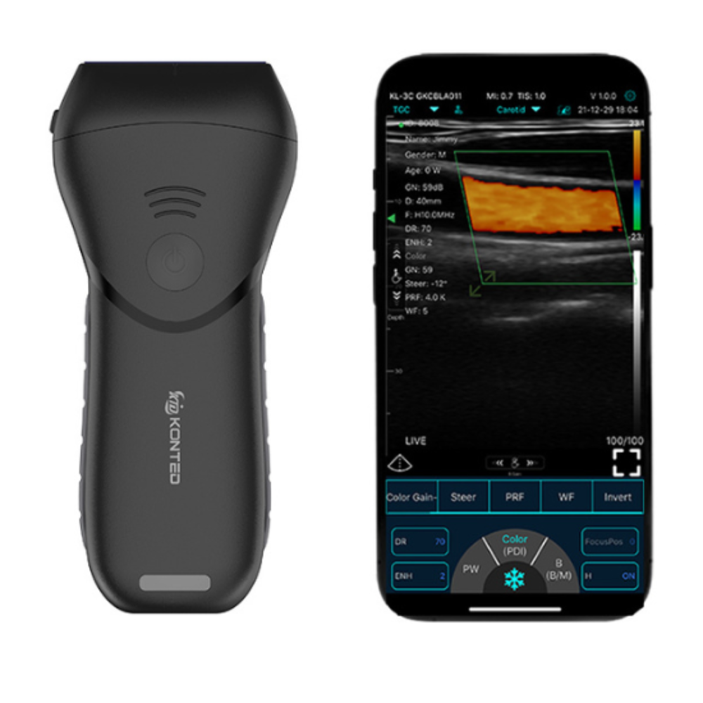
Superficial Ultrasound Linear Probe
The Superficial Linear Probe is designed for high-resolution imaging of structures located close to the surface of the body. Its high-frequency technology provides exceptional detail, making it ideal for visualising muscles, tendons, ligaments, nerves, blood vessels, thyroid tissue, soft tissue structures, and other superficial anatomy. Whether used for diagnosis, treatment planning, image-guided procedures, or education, it delivers the clarity clinicians need to make more informed decisions at the point of care.
As healthcare continues to move towards faster, more accessible, and patient-centred care, portable ultrasound is becoming an increasingly valuable tool across a wide range of clinical settings. From physiotherapy and sports medicine to pain management, aesthetics, primary care, vascular assessment, and medical education, clinicians are using high-frequency linear ultrasound to improve confidence, enhance patient communication, and support more precise clinical interventions.
The portability of the MX Pro allows clinicians to bring diagnostic imaging directly to the patient rather than relying solely on referrals to imaging departments. This can help streamline clinical workflows, reduce delays, support earlier decision-making, and improve the overall patient experience. Combined with advanced imaging modes including Colour Doppler, Power Doppler, Pulsed Wave Doppler, and other diagnostic capabilities, the system provides a versatile solution for everyday clinical practice.
Below are some of the most common clinical applications where the Superficial Linear Probe can support assessment, procedural guidance, monitoring, and education. While these examples demonstrate the versatility of the probe, ultrasound should always be used within the clinician's scope of practice, level of training, and in accordance with relevant professional guidance.
1. Musculoskeletal (MSK) Assessments
Ideal for imaging muscles, tendons, ligaments, joints, bursae, and superficial soft tissue structures. Useful for physiotherapy, sports medicine, rehabilitation, orthopaedics, and pain clinics when assessing injuries or guiding treatment plans.
2. Guided Injections & Pain Management
Supports real-time visualisation during injections, helping clinicians guide needles with greater precision. Suitable for joint injections, tendon sheath injections, trigger point procedures, nerve-related pain treatments, and regenerative medicine applications.
3. Vascular Imaging & Blood Flow Assessment
With Colour Doppler, PW, and PDI modes, the MX Pro can assess superficial blood vessels and blood flow. Useful for vascular mapping, circulation assessment, vein localisation, and treatment planning.
4. Nerve Imaging & Regional Procedures
Helps visualise superficial nerves and surrounding tissue structures, supporting nerve blocks, pain management procedures, and anatomy-guided interventions.
5. Physiotherapy & Sports Injury Clinics
Portable design makes it ideal for private physiotherapy clinics, sports therapists, and mobile practitioners who need fast imaging for tendons, strains, soft tissue injuries, and rehabilitation monitoring.
6. Thyroid & Neck Scanning
Suitable for superficial neck structures including thyroid imaging, lymph nodes, and surrounding tissues. Can assist with screening, monitoring, and referral decisions.
7. Breast & Small Parts Imaging
Useful for superficial breast tissue, lymph nodes, and small parts examinations where high-frequency detail is beneficial.
8. Dermatology & Soft Tissue Assessment
Can be used to assess cysts, nodules, swelling, superficial lesions, and soft tissue irregularities. Helpful in dermatology, minor procedures, and primary care settings.
9. Point-of-Care Ultrasound (POCUS)
Its wireless portable design makes it ideal for rapid bedside or chairside assessments in clinics, urgent care, community healthcare, and private practice settings.
10. Education & Training
Excellent for teaching anatomy, ultrasound-guided injections, vascular access, MSK scanning, and practical clinical skills in universities, academies, and training centres.
ABOUT US
Oras Medical is a specialist clinical adoption provider helping professionals, clinics, hospitals, GPs and care teams implement portable medical ultrasound solutions across the UK.
Oras Medical is a trading name of Oras Marketing Limited, Registered in England & Wales with registration number 12935403. VAT No. GB498618427. Registered address: Eastlands Court Business Centre, St. Peters Road, Rugby, Warwickshire, CV21 3QP. Email: admin@orasmedical.com, Phone: +44 (0) 1788 270 191.
SOLUTIONS
PRODUCTS
USEFUL INFO
PAYMENT INFO

Copyright 2026 Oras Medical - Privacy policy | Terms & Conditions | Disclaimer